Shoulder in Gout MRI images is a series of 3 shoulder gout MRI scans that I use in Gout in Shoulder.
Shoulder in Gout MRI images
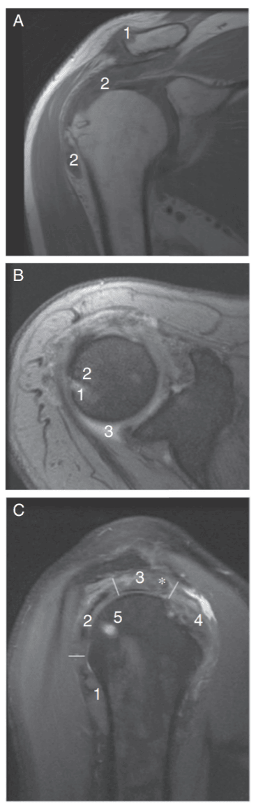
These images are from the Spanish gout case study featured in my Gout in Shoulder article. So, I have translated the original caption using Google Translate. But note my translation of intrathendinous which I believe is best translated as intratendinous (within tendon part of the muscle):
A) MRI image of the right shoulder in coronal acquisition in a sequence enhanced in T1. In an image superimposable to the X-ray of Figure 1B, the tophi are seen as hypointense soft tissue masses in T1 at the upper edge of the distal clavicular end (1) and in the subacromial deltoid bursa (2).
B) Image in axial acquisition T2 gradient echo. MRI reveals a lesion compatible with gouty tophus (1) in the glenohumeral articular rim not clearly visible on plain radiography. It is hyperintense in T2-weighted sequence, interrupts the bone cortex and has a very hypointense rim due to bone sclerosis (2). A discrete synovial effusion is associated (3).
C) Image in sagittal acquisition in proton density that allows seeing the section of the tendons that form in rotator cuff: minor round (1), infraspinatus (2), supraspinatus (3) and subscapular (4). The supraspinatus is especially unstructured due to the presence of a nodule suggestive of an intratendinous (within tendon part of the muscle) tophus (*). As in the T2-weighted sequence, the bony tophus is hyperintense (5).
Leave Shoulder in Gout MRI images to read Gout in Shoulder.
Please give your feedback
Did this page help you? If yes, please consider a small donation. Your donations help keep GoutPal's gout support services free for everyone.
If not, please tell me how I can improve it to help you more.
- YouTube
- The gout forums.